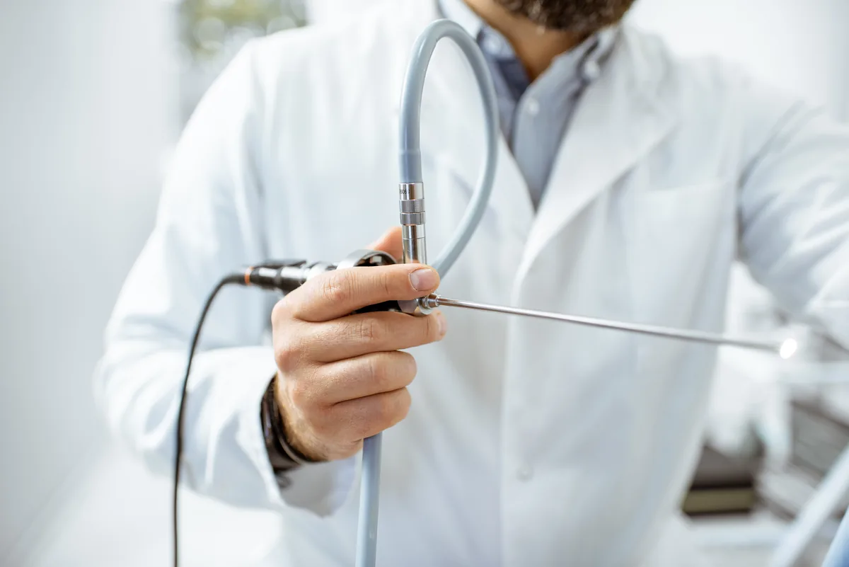
Treatment of Kidney Stones with Flexible Ureteroscope
Access is achieved through urinary canal called urethra, without making an incision. A thin guide wire is placed, a sheath called “access sheet” is placed to the ureter over the wire and the flexible ureteroscope is advanced to the kidney through it. After the stone is visualized with the camera, laser wire, measuring 230 microns in thickness, is advanced through flexible ureteroscope, direct contact with the stone is established and the stone is fragmented. Fragmentation of the stone is maintained until fragments are reduced to sand size.
Kidney stones have been the most painful condition among urologic diseases for centuries. The researchers found that kidney stones were present in Egyptian mummies, 7000 years ago. Urinary tract stones imply the third disease that affects urinary tracts following prostate pathologies and urinary tract infections. Millions of individuals present to urologists or emergency physicians due to kidney stones annually. In most cases, kidney stones may pass without intervention while causing mild or severe pain. However, the stone may persists depending on its size and shape and characteristics of the patient’s canal structure. A stone, measuring 5 mm in diameter, has 50% chance of passing. The chance of passing decreases as the size of the stone increases.
While open surgery was used for kidney stones before 1980, the rate of this method decreased to 1-5% of the total cases recently. Currently, most of the stones are treated with ESWL “Extracorporeal shock wave lithotripsy” and endourological “endoscopic closed” methods. Those which are less invasive started to stand out among these methods, which are preferred more frequently in recent years.
Technological improvements made on devices and equipment used in flexible ureterorenoscopy within the last decade placed the retrograde intrarenal surgery to an essential position among minimally invasive methods.
How is flexible ureteroscopy performed?
Access is achieved through urinary canal called urethra, without making an incision. A thin guide wire is placed, sheath called “access sheet” is placed to the ureter over the wire and the flexible ureteroscope is advanced to the kidney through it. After the stone is visualized with the camera, laser wire, measuring 230 microns in thickness, is advanced through flexible ureteroscope, direct contact with the stone is established and the stone is fragmented. Kırılan parçalar kum tanesi haline gelene kadar taş kırılır.
Why is Flexible Ureteroscopic treatment superior to ESWL method?
ESWL is a method which has been used successfully in treatment of kidney stones for long years. However, flexible ureteroscope is a more effective solution considering long durations of kidney stone treatment sessions (1-4 sessions), unsuccessful procedures especially regarding kidney stones in certain areas (success rate is 40-60 % for stones in lower section of kidneys) and undesired side effects following ESWL such as residual stone fragments. Today, flexible ureteroscopy is the first option for stones that cannot be fragmented with ESWL. Moreover, it is the primary option for stones measuring less than 1.5 cm in lower parts of kidneys due to unsuccessful results of ESWL.
What are the fields of use of Flexible Ureteroscopy other than treatment of stones?
The method can be used for investigation of bleeding in urinary tracts and with diagnostic purposes for patients with positive upper urinary system pathology (suspected tumor in ureter or kidney). However, currently, the most common and successful field of use is kidney stones.
What are the advantages of kidney stone treatment with flexible ureteroscope?
The best advantage of the method is the high success rate. (90-96)
On the contrary to methods such as percutaneous and open surgery, no incision scars remain in the body as the kidney is accessed through natural routes.
The studies showed that the method does not cause tissue or function loss in kidney.
It does not cause bleeding. Therefore, availability of the procedure for patients with tendency to bleed and patients who use anticoagulant agents provides a great advantage.
The patient can be discharged on the same day or the day after.
It can be performed for treatment of patients with congenital kidney anomaly such as pelvic and horseshoe kidney. It is one of the most effective and easy methods used for this patient group.
One of the major advantages is the ability to intervene the stones both in ureter and kidney simultaneously. A kidney stone can be incidentally found at ipsilateral side, while ureteral stone is treated with ureteroscope. In such cases the kidney stone in the same side can be removed after the stone in ureter is extracted, which allows eliminating all of the stones.
Stones in bilateral kidneys can be removed in the same flexible ureteroscopy session for patients who require treatment for stones in both kidneys.
ESWL cannot be performed for patients with excessive body weight and percutaneous ureteroscopy is a challenging procedure which may lead to various side effectsTherefore flexible ureteroscopy is considered as the primary option for treatment of stones in such patients.
Is it possible to treat stones in both kidneys at the same time?
This is one of the major advantages of using flexible ureteroscopy in treatment of stones. The stones in right and left kidneys can be treated within the same session if the stone load is not significantly excessive.
Can it be performed for the stones in upper sections of ureter?
Stones in upper end of ureter are among the type of stones where flexible ureteroscopy is most successful. The success of ESWL is relatively lower in treatment of these stones and correction of obstruction caused by these stones cannot be achieved immediately. The quickest and most effective treatment method in this situation is the eradication of the stone using Ho laser and Flexible ureteroscopy.
One of the major problems in use of rigid ureteroscopy for treatment of stones in middle, lower or upper sections of ureter is the possibility of migration of the stone to the kidney. During the period when flexible ureteroscopy was not used, a catheter named double j were placed to the area before procedures were terminated and the treatment of the stones were postponed to be completed with other methods in cases where stone migrated to the kidney. With flexible ureteroscopy method, kidney can be accessed to fragment the stone in cases of migration of stone to the kidney and stone can be treated in the same session.
How long is the hospitalization period?
Patients are able to return to their normal activities quickly as no bleeding occurs during the procedure, no incisions are made and no holes are opened. Patients can be discharged on the same day.
Is it possible to perform flexible ureteroscopy for patients with excessive weight?
Yes, ESWL treatment cannot be used for these patients in most cases and percutaneous method is challenging. Flexible ureteroscopy is an excellent option for these patients. Various international scientific studies point that flexible ureteroscopy should be the primary option as complication and blood transfusion rates are high in percutaneous applications.
The content on this page has been prepared by Güven Hospital solely for general informational purposes. The information provided does not replace diagnosis or treatment. For evaluation and treatment planning related to your health condition, please consult your physician.