EBUS (Endobronchial Ultrasound)
Bronchial and Mediastinal Evaluation
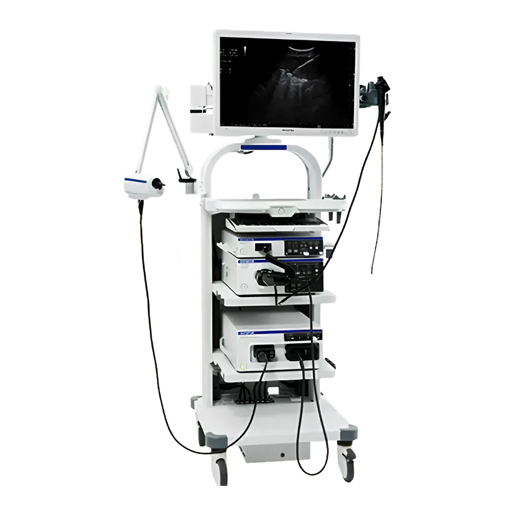
Endobronchial Ultrasound (EBUS) is an advanced imaging and diagnostic technique used for the evaluation of lymph nodes and masses within the lungs and mediastinum. By combining bronchoscopy with real-time ultrasound imaging, EBUS plays a critical role in the diagnosis and staging of lung cancer. As a minimally invasive procedure, it provides highly accurate diagnostic information without the need for surgical intervention.
Technological Infrastructure and Procedure
EBUS is performed using a specialized bronchoscope equipped with an integrated ultrasound probe at its tip. During the procedure, the bronchoscope is advanced through the mouth or nose into the tracheobronchial tree, allowing real-time ultrasound visualization of structures beyond the bronchial wall.
Simultaneously, tissue sampling can be performed using Endobronchial Ultrasound-Guided Transbronchial Needle Aspiration (EBUS-TBNA). This technique enables direct sampling of suspicious lymph nodes or masses under real-time ultrasound guidance. The procedure can be carried out under general anesthesia or moderate (conscious) sedation and is typically completed within a short period of time.
Advantages
- Minimally invasive and can be performed safely without the need for surgical intervention.
- Provides highly accurate and reliable results for lung cancer staging.
- Real-time ultrasound guidance ensures precise targeting of lymph nodes and lesions during biopsy.
- Short recovery time and a low complication rate contribute to enhanced patient comfort and safety.
- Diagnosis and staging can often be completed during the same procedure, facilitating faster treatment planning.
Clinical Applications
EBUS is widely utilized in pulmonology and thoracic oncology for a variety of diagnostic and staging purposes:
- Lung Cancer: Diagnosis, staging, and treatment planning.
- Lymphoma: Evaluation and sampling of mediastinal lymph nodes.
- Tuberculosis and Sarcoidosis: Tissue acquisition for the diagnosis of granulomatous diseases.
- Metastatic Disease: Assessment of pulmonary and mediastinal involvement from primary malignancies located elsewhere in the body.
- Mediastinal Masses: A reliable diagnostic tool for the evaluation and differential diagnosis of mediastinal lesions.

Ваше здоровье
В руках доверия
Новое мобильное приложение Güven Health Group уже доступно!
Скачивайте наше приложение с легкостью! Выберите ссылку, соответствующую вашей операционной системе, и сделайте свою жизнь проще прямо сейчас.